

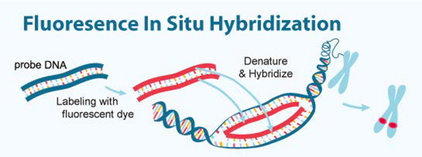
Real-time Fluorescence in Situ Hybridization for Detection of Pathogens
Professor: Abhinav Bhushan (abhushan@iit.edu)
Department: Biomedical Engineering
Brief Description: Fluorescence in situ hybridization (FISH) is widely used for the detection of specific nucleic acid sequences in cells, tissue sections, and whole organisms. FISH reactions are typically performed using a bench-top assay by pipetting the hybridization reagents onto the cytological sample, in which the transport of probes into the cell is primarily diffusion-based, which leads to typical incubation times of tens of hours. We have previously carried out real-time detection of proteins and small molecules using continuous flow kinetics in a microfluidic device. The goal of this project will be to design a microfluidic device and sets of probes that can be used for real-time detection of pathogenic strains using in situ hybridization.Preferred student skills: Major in Biology, Chemistry, or Biomedical Engineering.
Expected student outcomes: The student will specifically learn about chemical and biological assay development, bacterial pathogens, and microfluidics. During this process, the student will achieve:
- an ability to develop and conduct appropriate experimentation, analyze and interpret data, and use engineering judgment to draw conclusions
- an ability to communicate effectively with a range of audiences
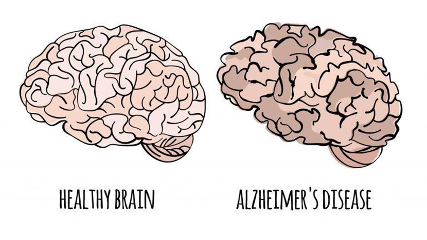
Ultrasensitive protein detection in Alzheimer's Disease
Professor: Abhinav Bhushan (abhushan@iit.edu)
Department: Biomedical Engineering
Brief Description: Millions of people worldwide are suffering from Alzheimer’s Disease (AD), and unfortunately, there is currently no cure available and no way to prevent the progression of damage. There are treatments available to manage the symptoms of the disease, however, since damage can initiate decades before the onset of clinical symptoms, early diagnosis is the key. However, there are several limitations in imaging the protein biomarkers including very poor specificity in differentiating AD patients from control subjects. Ideally, measuring blood-based biomarkers would be desired but the sensitivity and specificity of current assays carried out in samples containing serum is very poor. The goal of this research is to explore the development of an ultrasensitive assay for detection of proteins, based on work published in the Bhushan lab, which will be applied towards AD.
Preferred student skills: Major in Biology or Biomedical Engineering.
Expected student outcomes: The student will specifically learn about biological assay development, statistics, and Alzheimer’s disease. During this process, the student will achieve:
- an ability to develop and conduct appropriate experimentation, analyze and interpret data, and use engineering judgment to draw conclusions
- an ability to communicate effectively with a range of audiences
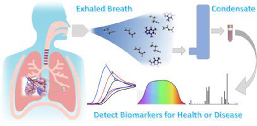
Development of sensors for exhaled breath
Professor: Abhinav Bhushan (abhushan@iit.edu)
Department: Biomedical Engineering
Brief Description: Current methods for detection of infections such as COVD-19 is carried out by reverse-transcription polymerase chain reaction analysis. Although this method is highly specific, the long assay time causes delay in the analysis which limits practical use such as real-time assessment of infection status. Such a technique could be mobilized to screen large numbers of people, and can immediately identify persons who require confirmatory testing. One promising screening method is to use exhaled breath, which involves analysis of exhaled volatile organic compounds that are endogenously formed as metabolic byproducts. It is known that infectious pathogens can alter the composition of breath compounds, therefore, unique volatile compounds in the exhaled breath can be used for early and rapid diagnosis. However, the promise of using volatile compounds for diagnosis has not reached its potential. One of the primary reasons for this is that current sensors have very poor specificity towards volatile compounds.
Preferred student skills: Major in Biology, Chemistry, or Biomedical Engineering.
Expected student outcomes: The student will specifically learn about sensor technologies, volatile organic compounds, and experimental design. During this process, the student will achieve:
- an ability to carry out literature review
- an ability to develop and conduct appropriate experimentation, analyze and interpret data,
- an ability to communicate effectively with a range of audiences
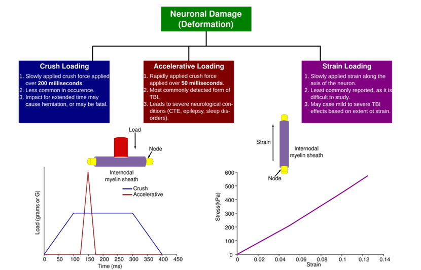
Determination of the biomechanical properties of mammalian neuronal tissues at nanoscopic and mesoscopic scales
Professor: Joseph Orgel (orgel@iit.edu)
Department: Biology
Brief Description: It is well understood that nerves, nerve bundles, individual neurons are cells and tissues of significant influence on the mammalian body. Within the key functional structures of a subset of neurons is the packing of myelin layers of both central and peripheral nervous systems. Myelin is used by myelinated nerves to accelerate the transmission speed of nervous signals. When myelin structure is altered, such as in some (but certainly not all) instances of mechanical loading to the organism, the ability of the myelinated nerves to conduct signal is altered or impeded entirely. This can lead to severe developmental, behavioral, or otherwise biological impediments to the normal function of the effected tissues and the organism.
When substantial changes to myelin occur, they are relativity apparent in testing and clinical imaging and severe biological impairment is likely, such as in Traumatic Brain Injury (TBI). However, there can be ‘invisible’ changes to myelin, not readily detectable in living specimens or even postmortem, that lead to mild, moderate or severe biological impairments. It is reasonable to suppose, that mild or moderate mechanical loading leads to minute changes in myelin that are notoriously difficult to detect due to their small scale, but lead to significant biological and behavioral changes. This study is intended to bridge this impasse and identify what type of loading at what extent leads to what kind of permanent deformation of myelin structure.
Determination of tissue response to mechanical damage (stress and strain) is crucial to understanding and developing dose-response models of various tissues. These data are of high value to developing high-fidelity computational models of tissue and constituent elements.
This study will provide basic biomechanical data that has a wide range of applications in the characterization of living matter (in this case neurons) but especially so in the interpretation of traumatic injury computations and design of protective equipment. With major innovations in the field of simulated surgery for training of surgeons, these data will provide significant improvements on tissue definition, boundaries and biomechanical responses to stress, strain and impact during surgery, thus improving feedback fidelity to train surgeons better.
This study uses methods previously developed by the Orgel research group to establish a mechanical dose-response model for animal and human optic nerves. Optic nerves will be stretched and compressed at various rates to study damage using microscopy (at IIT) and X-ray diffraction (BioCAT, APS, ANL). The study will utilize animal and cadaveric human tissues to establish these mechanical dose-response properties.
Preferred student skills: Anatomical dissection of animal and human tissues, general laboratory etiquette (cleanliness and safety), motivated and enthusiastic to learn new techniques, team player.
Expected student outcomes: Enhanced dissection and sample preparation techniques for ex vivo testing of tissues, mechanical dose-response data analyses using ImageJ and Python, basics of X-ray diffraction data analyses using MuscleX and Python.
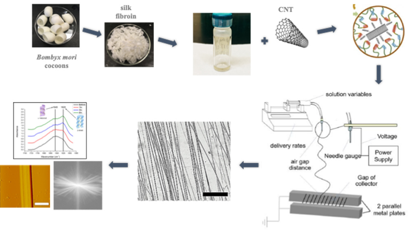
Optimization of electrospinning conditions to achieve aligned silk composite nanofibers
Professor: Rong Wang (wangr@iit.edu)
Department: Chemistry
Brief Description: Silk fibroin, a structural protein of the Bombyx mori cocoons, has been widely used in biomedical applications because of its superior mechanical properties, biocompatibility, low degradation rate and ease of processing. Our previous work has shown that electrospinning technique can be employed to generate aligned silk protein fibers, mimicking the structure and dimension of matrix proteins in the extracellular matrix of connective tissues, thus, can be used as tissue engineering scaffolds. The addition of functionalized carbon nanotube (CNT) to the silk protein was shown to reinforce the strength of the scaffolds and render the fibers electrical conductivity. However, the addition of CNT also affected the fiber dimension, alignment, uniformity, stability, biocompatibility and cell adhesion. In this project, we aim to optimize the electrospinning conditions to achieve silk composite fibers with desirable properties.
Preferred student skills: Passionate about research, good hands-on skills.
Expected student outcomes: The student will receive trainings in (1) extracting and purifying silk fibroin from silk cocoons; (2) surface functionalization of CNT; (3) generating silk and silk composite nanofibers by electrospinning; (4) fiber characterization and data analyses.
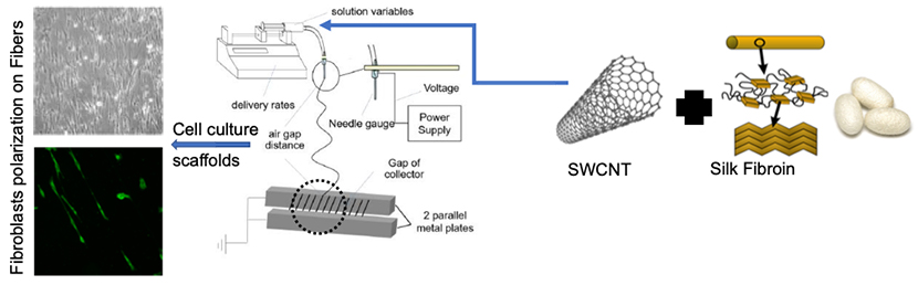
Modulation effect of silk-CNT fibers on cell-matrix interaction
Professor: Rong Wang (wangr@iit.edu)
Department: Chemistry
Brief Description: Functional biopolymer scaffolds are in high demand for tissue regeneration. A biocomposite scaffold uniquely combines the chemical and physical properties of materials derived from nature and synthetic systems to achieve the functionality that uniquely meets the need. We have generated silk-CNT composite fibers by electrospinning, which are mechanically strong, electrically conductive and unidirectionally aligned. When fibroblasts were cultured on the fibrous matrix, they polarized in the direction of fibril alignment due to the anisotropic deposition of focal adhesions and the enhanced cell contraction associated with the increased cytoskeletal tension induced by the stiffer silk-CNT fibers. The activated fibroblasts could produce collagen at an increased level, which is critical in tissue repair. We found fibril density, cell seeding density and the source of the fibroblasts play important roles in fibroblast activation. In this project, we aim to vary these conditions and examine the modulation effect of the silk-CNT fibers on cell-matric interactions.
Expected student skills: Passionate about research, good hands-on skills, 2+ years labs.
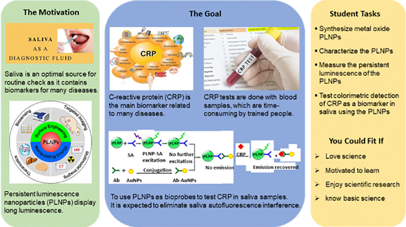
Colorimetric detection of biomarkers in saliva using luminescence nanoparticles
Professor: Yuanbing Mao (ymao17@iit.edu)
Department: Chemistry
Brief Description: Persistent luminescence nanoparticles (PLNPs) can remain luminescent for hours after cessation of excitation. They have emerged as important materials in biomedicine due to their special ability to eliminate tissue autofluorescence. In this study, we will explore the colorimetric detection of biomarkers in saliva using metal oxide PLNPs as bioprobes.
Specifically, we will apply metal oxide PLNPs as bioprobes to determine the concentrations of C-reactive protein (CRP) for periodontitis diagnosis. CRP is an acute-phase reactant that is found at elevated levels in the whole saliva of patients who have periodontal disease. It is also the main biomarker related to cardiovascular risk and heart attack occurrence. Since the PLNP bioprobes display long persistent luminescence, saliva autofluorescence interference can be eliminated.
It is expected that the developed PLNPs possess tunable size and desirable persistent luminescence as ideal probes for eliminating autofluorescence interference in biosensing. This work is valuable in research areas such as studying the functions of biomolecules and monitoring of molecular/cellular networks in their native contexts.
Preferred student skills: Motivated and dedicated, enjoy scientific research with basic science knowledge.
Expected student outcomes: Synthesis and characterization of metal oxide nanoparticles. Test of persistent luminescence of the nanoparticles before and after interacted with CRP.
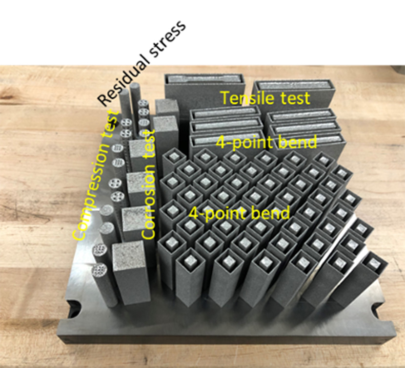
3D-printing of cost-effective titanium alloys: manufacturing, characterization, and biocompatibility assessments
Professor: Amir Mostafaei (mostafaei@iit.edu)
Department: Mechanical, Materials, and Aerospace Engineering
Brief Description: The proposed project aims to thoroughly assess the role of the powder characteristics and laser processing of non-spherical powders to understand physical phenomena occurring during powder spreading dynamics and laser melting via advanced characterization tools to minimize two main common issues associate with laser powder bed fusion machines including pores formation and time of production. Mechanical properties such as tensile and compressive strengths, ductility, and fatigue will be assessed on the as fabricated and post-processed additive manufacturing (AM) parts and results will be correlated to microstructure, defects and surface finish. In addition, we evaluate biocompatibility as a function of the process condition of AM parts.
This research merges disciplines including materials science, advanced materials processing, and biomedical engineering. By both the manufacturing efficiency and the cost-effectiveness of the AM biomedical components made using high-quality, cost-effective powder, the knowledge gained from these investigations will provide significant opportunities for the biomedical industry.
Preferred student skills: Motivated, enjoy manufacturing and characterization, know basics of materials science.
Expected student outcomes: Operating laser powder bed fusion machine, feedstock and 3D printed part characterizations, process optimization, data analysis.
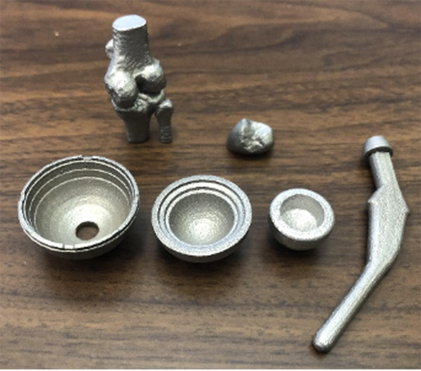
Development of bioimplants using advanced manufacturing material
Professor: Amir Mostafaei (mostafaei@iit.edu)
Department: Mechanical, Materials, and Aerospace Engineering
Brief Description: Over the past decades, 3D printing or additive manufacturing (AM) has evolved substantially and has proven significant potential in biomedical applications. 3D printing is a material-oriented manufacturing technology, since the solidification mechanism, architecture resolution, post-treatment process, and functional application are based on the materials to be manufactured. However, there are limited numbers of biomaterials that can be processed using AM to fabricate bioimplants. We will explore different biomaterials such as titanium alloys, stainless steels and cobalt-chromium based alloys and fabricate complex parts such as hip and joints, dental crown, partial denture framework, and craniofacial reconstruction. Microstructure, defects, mechanical properties, electrochemical behavior, and biocompatibility of parts will be evaluated and compared to conventionally manufactured alloys through casting and metal forming processes.
Preferred student skills: Motivated, enjoy manufacturing and characterization, know basics of materials science.
Expected student outcomes: Operating 3D printers, process optimization, design, materials characterizations, mechanical testing.
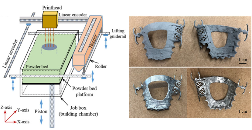
Understanding role of powder characteristics on part density and surface roughness of binder jet 3D printed biomaterials
Professor: Amir Mostafaei (mostafaei@iit.edu)
Department: Mechanical, Materials, and Aerospace Engineering
Brief Description: In recent decades, substantial progress has been made in the understanding, development, and utilization of additive manufacturing processes. Binder jet 3D printing (BJ3DP), a non-beam based additive manufacturing (AM) method, refers to the technology in which powdered material is deposited layer-by-layer and selectively joined in each layer with a thermosetting binder, then followed by post-processing to consolidate parts. In the proposed work, we aim to advance the state of technology on binder jetting of ultra-fine powder (stainless steel 316L) incorporated with an innovative manufacturing system of using ultrasonic powder dispenser to enhance powder packing density and minimize powder bed defects. Further, results will be compared with printed parts using coarse metal powders. Participant student will be trained out designing, 3D printing and characterization aspects of the additively manufactured implants and biostructure parts.
Preferred student skills: Motivated, enjoy manufacturing and characterization, know basics of materials science.
Expected student outcomes: Operating 3D printers, process optimization, design, materials characterizations, mechanical testing.